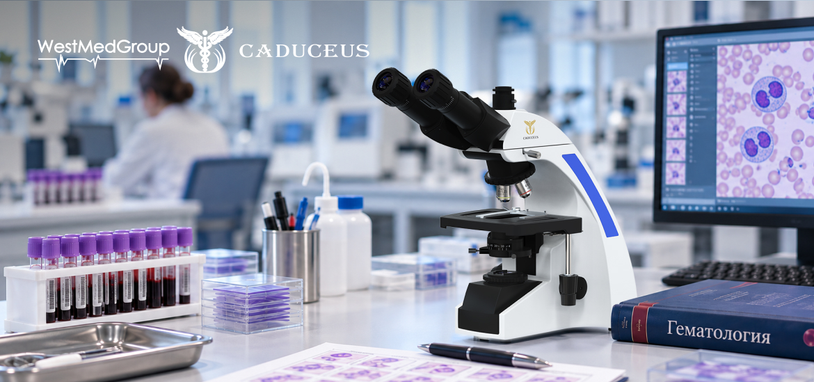
A microscope in hematology is more than a magnification tool — it is an essential part of accurate morphological diagnostics, where image quality can make all the difference. The article explores the key factors to consider when selecting equipment, helping specialists examine the smallest cellular changes and turn invisible details into the basis for reliable conclusions.
When examining blood smears, even minor differences in cell morphology can be critical for establishing a diagnosis. Therefore, image quality remains one of the key factors affecting the accuracy of morphological assessment and the subsequent interpretation of results.
Modern hematological diagnostics combine automated analyzers with microscopic examination. While automated systems provide high sample-processing speed, a microscope enables detailed examination of blood cell composition, clarification of questionable results, and confirmation of changes that require professional assessment. For this reason, selecting the right laboratory instrument is an important part of ensuring efficient workflow.

In a hematology laboratory, a microscope is used for a wide range of diagnostic tasks. It enables specialists to identify atypical cell forms, assess changes characteristic of various blood disorders, and confirm results obtained using automated analyzers.
When selecting equipment, it is important to consider not only magnification, but also the quality of the optical system, resolution, illumination characteristics, focusing convenience, and the ergonomics of the design. For many laboratories, the availability of photo and video documentation of specimens, compatibility with digital cameras, and integration with additional equipment are becoming increasingly important. These features make it possible to conduct remote consultations and use captured images for educational purposes.
Among modern solutions that meet these requirements are CADUCEUS laboratory microscopes. Their trinocular design allows digital cameras and additional equipment to be connected, facilitating photo and video documentation of specimens, recording of examination results, and the use of captured materials for training and consultations. High-quality optics, adjustable LED illumination, and the ability to configure illumination according to the Köhler illumination method provide the clear and high-contrast images required for morphological examinations.
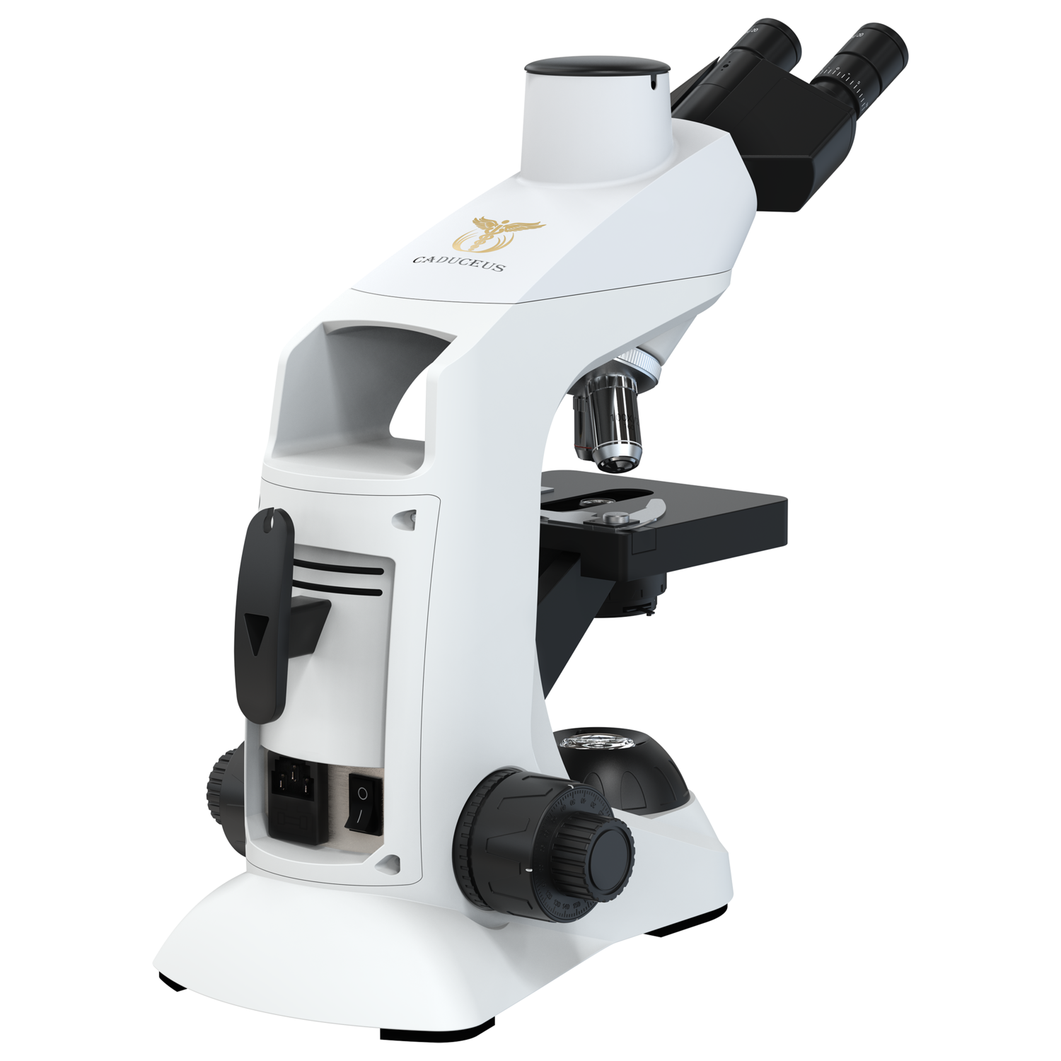
Furthermore, the CADUCEUS product range includes a variety of models, making it possible to select equipment based on the specific needs of the laboratory, examination workload, and functional requirements. By combining modern optical solutions, an ergonomic design, and digital integration, these microscopes are suitable both for routine laboratory practice and for tasks involving result documentation and educational activities.
Thus, the choice of microscope for a hematology laboratory directly affects the quality of morphological diagnostics and the efficiency of laboratory specialists. When evaluating equipment, it is important to consider not only its technical specifications, but also ease of use, digital imaging capabilities, and suitability for the laboratory’s specific requirements. A properly selected microscope helps ensure reliable examination results, optimize the daily workflow of specialists, and maintain a high standard of laboratory diagnostics.